smartCONCEPT
smartDESIGN
Study Title
Virtually Wall-Less versus Standard Thin-Wall Venous Canula in the Minimally Invasive Mitral Valve Surgery: Single-Center Experience
Fabricio Ceresa et al
Design
Retrospective
Observational
Objective
“Benefit assessment of using a virtually wall-less canula in comparison with the standard thin-wall canula in clinical practice”
Patients
65 consecutive elective patients undergoing minimal invasive mitral valve surgery
smartcanula compared to standard thin wall cannula
During CPB, the mean number of red blood cell units transfused was significantly higher in standard cannula group than in the smartcanula group. This may be due to the greater haemodilution required to maintain the estimated target flow when we used the thin-walled cannula.
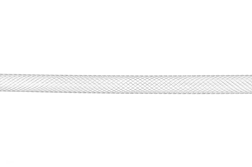
The greater mean cross-sectional diameter of the self-expanding design is attributable to the collapse insertion and expanding in situ approach. This has resulted in an intravascular diameter greater than the access channel diameter. It is a well-known fact that this outcome is unattainable with traditional rectilinear cannulas.
Another salient point pertains to the substantially reduced negative pressure necessitated for a full flow, a characteristic that is inherently embodied by the smartcanula, in comparison to the standard thin-wall percutaneous cannulas.
In larger patients, the smartcanula enables a more bloodless surgical field in comparison to the conventional cannula. This results in a considerable reduction in aortic cross-clamp time and a more satisfied cardiac surgeon.
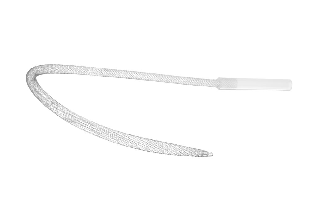
Inadequate venous drainage during MICS not only causes discomfort for the surgeon but also does not allow the estimated target flow to be reached, which is defined as 2.4 lt/min/m2. For these reasons, the virtually wall-less cannula was created as a temporary caval stent to maximize venous drainage, preventing the caval axis from collapsing despite the negative pressure applied by the augmentation through the venous line.
This design, in the same manner as a grid, allows direct venous drainage at all levels of caval affluent vessels, particularly the renal and hepatic veins.Moreover, the self-expanding characteristic of the wall avoids the caval injury caused by the oversizing of the cannula.
smartINSERTION
Study Title
The Smart Canula: A new tool for remote access perfusion in limited access cardiac surgery
Ludwig von Segesser et al
Design
Observational
Objective
“Assessing the performance of the smartcanula in a small series of patients undergoing redo procedures”
Patients
5 patients; 2 males and 3 females; mean age 62 ± 7 years; Undergoing redo procedures of the aortic valve (2) and ascending aorta (2); thoraco-abdominal aorta
The smartCANULATION Concept
The smartCANULATION concept is designed to take advantage of the geometric configuration of the venous vasculature when remotely cannulating through a smaller access vessel.
The smartcanula principle of collapsed cannula insertion and expansion in situ provides an efficient new solution for an old problem, which is inadequate venous drainage during perfusion.
The insertion of a venous cannula in collapsed state, which can be expanded within the target vessel, can almost match the diameters of the natural drainage vessels over a large proportion of its intravascular length.
As a matter of fact, 4 L/min can be achieved with a 20 F orifice for access, which is a fraction of the traditionally used orifice sizes for right atrial central cannulation.
smartUSE
Study Title
Single-Center Experience With a Self-Expandable Venous Canula During Minimally Invasive Cardiac Surgery (MICS)
Karel M. van Praet et al
Design
Retrospective data analysis
Objective
“Venous drainage is often problematic in minimally invasive cardiac surgery (MICS). The authors describe their experience with a self-expandable stent canula designed to optimize venous drainage.”
Patients
58 consecutive patients undergoing MICS for
• Mitral valve disease n = 40
• Left atrial myxoma n = 3
• Left ventricular outflow tract obstruction n = 1
• Aortic valve replacement n = 14
CPB Setup and Strategy
(1) Vacuum assisted venous drainage between -20 mmHg and -35 mmHg
(2) Target flow index reached 2.2 L/min/m² (at core temp of 34°C using a goal directed perfusion strategy aimed at minimum
The application of the self-expanding stent-like smartcanula is safe, effective, and reliable and simplified the procedure in minimally invasive MV and aortic valve surgery.
Drainage capacity is optimized, as the smartcanula takes advantage of the full size of the recipient vein.
Moreover, because this canula acts as a spring within the vein, the latter is maintained wide open by the canula itself, thereby limiting the venous collapse phenomenon.
smartPERFORMANCE
Study Title
Clinical experience in minimally invasive cardiac surgery with virtually wall-less venous cannulas
Ferrari et al; Innovations 2018;13:104–107
Design
Observational
Objective
„Inadequate peripheral venous drainage during minimally invasive cardiac surgery (MICS) is a challenge and cannot always be solved with increased vacuum or increased centrifugal pump speed. The present study was designed to assess the benefit of virtually wall-less transfemoral venous cannulas during MICS.“
Patients
10 consecutive patients undergoing MICS for Mitral (6), Aortic (3), and other (4) procedures
Patient characteristics 59 ± 10 years and 8 males, 2 females
smartCANULATION procedure
For a body size of 173 ± 11 cm and a body weight of 78 ± 26 kg, the calculated body surface area was 1.94 ± 0.32 m2.
As a result, the estimated target flow was 4.66 ± 0.78 l/min
whereas the achieved flow accounted for 98 ± 0.69 l/min (107% of target) at a vacuum level of 21.3 ± 16.4 mm Hg.
Once full flow on CPB was established, there was usually no further required manipulation and blood drainage remained stable throughout the operation.
Furthermore, the selected set-up allowed for excellent exposure and “dry” intracardiac surgical field.
We conclude that the virtually wall-less cannula designed for augmented venous drainage is a promising drainage device, which provides superior flow at reduced negative pressure, and thus, we expect also superior performance in routine clinical use.
smartPROCEDURE
Study Title
Virtually Wall-Less versus Standard Thin-Wall Venous Canula in the Minimally Invasive Mitral Valve Surgery: Single-Center Experience*
Fabricio Ceresa et al
Design
Retrospective
Observational
Objective
“Benefit assessment of using a virtually wall-less canula in comparison with the standard thin-wall canula in clinical practice”
Patients
65 consecutive elective patients undergoing
minimal invasive mitral valve surgery
smartCANULATION procedure
(1) Femoral vessels exposed at the level of the right groin
(2) Canulation with Seldinger technique after heparin administration (300 UI/Kg) and enlargement of the vein with progressive dilators
(3) Collapsed smartcanula is inserted over a guidewire and positioned at the tip at the origin of the inferior vena cava
(4) After mandrel removal the smartcanula expands to its full diameter (24Fr in this case)
We have been able to demonstrate that the performance of the virtually wall-less canula was superior compared to the standard thin wall canula
Better venous drainage due to the design of the canula
Even in larger patients the virtually wall-less canula allows a more bloodless field to be obtained, leading to a significant reduction in aortic cross-clamp time and major satisfaction on the part of the cardiac surgeon
smartDECANULATION
Study Title
Virtually Wall-Less versus Standard Thin-Wall Venous Canula in the Minimally Invasive Mitral Valve Surgery: Single-Center Experience
Fabricio Ceresa et al
Design
Retrospective
Observational
Objective
“Benefit assessment of using a virtually wall-less canula in comparison with the standard thin-wall canula in clinical practice”
Patients
65 consecutive elective patients undergoing
minimal invasive mitral valve surgery
The removal of the virtually wall-less cannula requires the pinching of the cannula itself at its exit from the femoral vein when pulling out the device. This maneuver allows blood loss to be minimized.
References
1. Pinon M, Usefulness of Self-Expanding Drainage Cannula in Venovenous Extracorporeal Membrane Oxygenation:
Tips, Tricks, and Results of an Early Experience; ASAIO Journal 68(2):p e22-e26, February 2022
2. Von Segesser L, Superior flow for bridge to live with self-expanding venous cannulas
European Journal of Cardio-Thoracic Surgery, Volume 36, Issue 4, October 2009, Pages 665–669
3. Van Praet KM, Single-Center experience with a self-expandable venous cannula during minimally invasive cardiac surgery
Innovations 2022, Vol. 00(0) 1–8
4. Krajinovic L, Intraoperative transesophageal echocardiography in cardiac surgery with a focus on mitral valve reconstruction
HTG 2024 38:37-51
5. Straub A, Erfahrungen mit der Smartcanula zur venösen Drainage im Langzeiteinsatz
Kardiotechnik 4/2011
6. Pinon M, Use of self expanding venous cannula in tricuspid reoperation
European Journal of Cardio-thoracic Surgery : 09 Jan 2015, 48(3):499-501
7. Ceresa F, Virtually Wall-Less versus Standard Thin-Wall Venous Cannula in the Minimally Invasive Mitral Valve Surgery:
Single-Center Experience; Medicina 2023, 59(7), 1221
8. Von Segesser L, A simple way to decompress the left ventricle during venoarterial bypass
Thorac Cardiovasc Surg. 2008 Sep;56(6):337-41
9. Strunina S, The peripheral cannulas in extracorporeal life support
Biomed Tech (Berl). 2019 Apr 24;64(2):127-133
10. Von Segesser L, The Smartcanula: a new tool for remote access perfusion in limited access cardiac surgery
The Heart Surgery Forum, 01 Jan 2005, 8(4):E241-5
11. Von Segesser L, New, virtually wall-less cannulas designed for augmented venous drainage in minimally invasive cardiac surgery
Innovations, Volume 11, Issue 4, 2016
12. Jegger D, A novel technique using echocardiography to evaluate venous cannula performance perioperatively in CPB cardiac surgery
European Journal of Cardio-Thoracic Surgery, Volume 29, Issue 4, April 2006, Pages 525–529
13. Belkoniene M, Performance increase in venous drainage for mini-invasive heart surgery: superiority of self-expanding cannulas
Innovations (Phila). 2014 Jul-Aug;9(4):297-301
14. Von Segesser L, Perfusion techniques during surgery oft he thoracic and thoraco-abdominal aorta: the veno-arterial bypass
Multimed Man Cardiothorac Surg. 2007 Jan1;2007(619):mmcts.2006.002535
15. Von Segesser L, Small access (30F) clinical central venous smart cannulation: is it adequate?
Interactive CardioVascular and Thoracic Surgery, Volume 5, Issue 5, October 2006, Pages 540–543